
Transmitted Light Microscopy
Transmitted light microscopy is the general term used for any type of microscopy where the light is transmitted from a source on the opposite side of the specimen to the objective lens. Usually, the light is passed through a condenser to focus it on the specimen to get maximum illumination. After the light passes through the specimen it goes through the objective lens to magnify the image of the sample and then to the oculars, where the enlarged image is viewed.
In order to get a usable image in the microscope, the specimen must be properly illuminated. The light path of the microscope must be correctly set up for each optical method and the components used for image generation. The condenser was invented to concentrate the light on the specimen in order to obtain a bright enough image to be useful. The microscope techniques requiring a transmitted light path include bright field, dark field, phase contrast, polarisation and differential interference contrast optics.
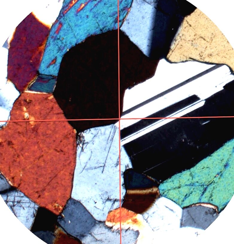
Polarised Light Microscopy
Polarised light microscopy uses plane-polarised light to analyse substances that are birefringent; i.e. matter that has two different refractive indices at right angles to one another like minerals. Normal, un-polarised, light can be thought of as many sine waves, each oscillating at any one of an infinite number of orientations (planes) around the central axis. Plane-polarised light, produced by a polar, only oscillates in one plane because the polar only transmits light in that plane. The polarised light microscope must be equipped with both a polarizer, positioned in the light path somewhere before the specimen, and an analyser (a second polarizer), placed in the optical pathway after the objective rear aperture. Image contrast arises from the interaction of plane-polarized light with a birefringent (or doubly-refracting) specimen to produce two individual wave components that are each polarized in mutually perpendicular planes. The velocities of these components are different and vary with the propagation direction through the specimen. After exiting the specimen, the light components become out of phase, but are recombined with constructive and destructive interference when they pass through the analyzer. Polarised light microscopy can be used to measure the amount of retardation that occurs in each direction and so give information about the molecular structure of the birefringent object (e.g. orientation).
Reflected Light Microscopy
Reflected light microscopy is used to examine opaque minerals (and other materials) in order to identify the mineral phases and determine the paragenetic relationships between the different mineral phases. The sample (polished thin section or polished button) is viewed using the reflected light microscope and can also be analysed using advanced x-ray and ion microprobe techniques. Bireflectance is an optical effect similar to pleochroism where the mineral appears to change in intensity as it is rotated while illuminated by plane polarised light. The polarisers are not crossed to observe bireflectance. Isotropic minerals (e.g, galena, pyrite) do not show any bireflectance (or pleochroism) when rotated in plane polarised light. Minerals which are pleochroic are also bireflectant. Care must be taken when observing bireflectance to follow these rules:
Sample is freshly polished and does not have any tarnish.
Illumination level is not too excessive (intensity changes the perceived relative intensity effect).
Minerals which are pleochroic (non-isotropic minerals) are also bireflectant.
Slicing granite to make thin sections.....